From Medicine to the Movies and Back
Dr Tom Shannon
The taking of what is believed to be the first photograph in 1826 by Joseph Nicéphore Niépce, gave scientists, anatomists and clinicians access to a new method to instantly record, analyse and characterise the gait of humans in health and when affected by a pathology. In 1872 Leland Stanford, a wealthy Californian race horse owner, wanted to prove the assertion that all a horse’s hooves are off the ground at the full gallop so hired a local photographer, Eadward Muybridge, to acquire the motion to answer the question. The un-intended outcome of this task set Muybridge on a new and life-long quest to study human and animal movement by creating his iconic but qualitative sequences of photographic plates. Inspired by the work of Étienne-Jules Marey, German scientist Christian Braune and his student Otto Fischer attempted to quantify human gait by applying Geissler tubes to body segments to simultaneously capture frontal and side views. For much of the latter part of the 19th and first half of the 20th century, exhaustive and tenacious work by these researchers and others was undertaken to extract meaning from the two dimensional projections of the limb segments within these early gait models with the objective to enlighten clinical understanding.
In the 1960’s, the opportunity to conduct routine three dimensional (3D) gait analyses was made possible through the hand digitisation of planar coordinates from two or more cine cameras with a photogrammetric reconstruction into 3D data points. The arrival of affordable computers and video camera in the 1970’s and 80’s were key additional innovations in helping to develop apparatus that could automatically reconstruct, label and track points in time and space. The commensurate development of 3D gait models and supporting software designed specifically for clinical applications combined with the new hardware to thrust the technology into the orthopaedic and biomechanics mainstreams with many subsequent advances further improving point and trajectory reconstruction accuracy, ease of use, availability and measurement consistency.
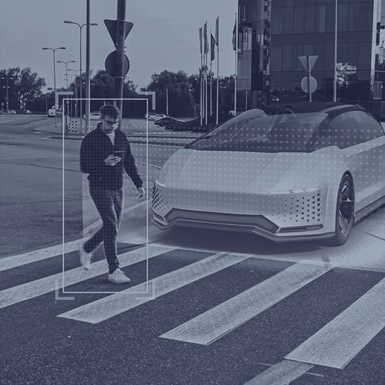
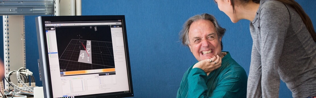
The most common application of movement analysis is to collect clinical information to aid in the understanding of the aetiology of presenting gait abnormalities and to assist in treatment decision-making [1]. Contemporary passive optical solutions are based on the acquisition and re-construction in three-dimensional space of the centres of small passive retro-reflective markers using multiple, synchronous cameras, optimised to produce data rather than a video image, and controlled by a computational device. In most life science applications the markers are placed directly onto the skin through palpating bony landmarks to derive quantitative information about the motion of anatomical points, segments and joint angles so facilitating the tracking and estimation of an underlying skeletal model. The reaction forces and torques produced between the patient and the ground can be simultaneously acquired to derive data including joint forces, moments and powers as can an appreciation of commensurate muscle electrical activity using specialised electrodes. The interpretation of resulting kinematic and kinetic data remains in the hands of interdisciplinary teams with substantial knowledge of normal and pathologic gait where characteristics are measured, abnormalities are identified, causes are postulated, and treatments are proposed [1].
The engineering problem to capture and track 3D points in space has been solved by a number of different approaches, with marker-less body part recognition possibly on the horizon, but clinical concerns remain over the loose association between the measurements of points on the skin surface and the underlying bone, commonly described as the soft tissue artefact [2] , a derivation of actual bone and joint locations so elegantly described by Dr David Sutherland as trying “…to measure the movements of a broomstick within a gunny sack?” [3]. Challenges remain to also reliably capture bony landmark axial rotation, in particular of the thigh [4]. Many researchers and gait laboratories have proposed techniques to move away from existing predictive approaches by instead discovering joint centres and axes functionally and fitting the data to an idealised joint model that also incorporates some form of soft tissue artefact compensation. This work continues and like the earlier innovations will gain wide acceptance only after it has been validated through close collaborative efforts between clinicians, scientists and engineers in laboratories, academia and industry.
The need to quantify sporting performance has resulted in the development of cameras with significant improvements in quantum efficiency that are now capable of working outside the laboratory to track fast and subtle motions with consistency for the measurement of both human and animal locomotion. The motion of the Mars Rover over desert terrain is an example of an outdoor application together with capturing the gait of kangaroos and even emus.
Since the release of the film Titanic in 1997 there has been an explosion of the use of motion capture to enhance films, games, music videos and television that have brought many stories to life, thought almost impossible before the ready availability of the emerging technology to the entertainment sector. With it has brought many challenges including the needs to capture motion from multiple characters simultaneously, in large volumes and with very high camera counts (243 in the case of the film Beowulf, 2007). The sector has also demanded improved subtlety in the measurement of the motion of hands, feet and facial expressions with a need to provide real-time reconstruction of the animated characters who may or may not be humanoid. All of this has been achieved and what is equally important is that the demands of each of the sectors has created a requirements synergy between them. The comprehensive understanding of anatomy and physiology in the life sciences has benefited the animators to create realistic characters, whilst their demands has directly improved how the technologies are applied and data are presented within the clinical gait laboratory.
- Davis RB, Õunpuu S, DeLuca PA, Romness MJ, Clinical Gait Analysis and Its Role in Treatment Decision-Making. MedGenMed 1(1), 1999 [formerly published in Medscape Orthopaedics & Sports Medicine eJournal 2(5), 1998]. Available at: http://www.medscape.com/viewarticle/440148
- Charlton IW, Tate P, Smyth P, Roren L. Repeatability of an optimised lower body model. Gait and Posture. 2004;20:213–221. doi: 10.1016/j.gaitpost.2003.09.004.
- Sutherland DH. The evolution of clinical gait analysis Part II Kinematics, Gait and Posture 16 (2002) pp 159–179.
- Lamoreux LW. Errors in thigh axial rotation measurements using skin mounted markers. 1991. In International Society of Biomechanics, The Department of Human Movement Studies, pp. 372–373.